
Enzyme Kinetics
A biological catalyst, enzymes act to accelerate a reaction without using up or changing themselves. There is just one type of reaction in which these enzymes work well along with just one type of reactant that is known as a substrate, or they just work with a small number of closely related compounds. Cells need enzymes for many biological reactions to occur as, without them, life would be impossible. An enzyme kineticist studies the rate at which enzymes react and the circumstances that influence those rates.Enzyme structure
Proteins, such as enzymes, are usually globular in their tertiary structure. There are reactants involved where there is an active site where the reaction takes place specifically the reaction they catalyze. An enzyme has a small cleft in its center where an amino acid structure binds substrates, forming a complex known as the enzyme-substrate complex (ES), which can be dissociated by a weak bond as soon as the reaction is complete. These amino acids are gathered by the rest of the enzyme, which acts as a scaffold.A specific substrate's shape complements the active site. Generally, there are two ways in which this interaction takes place:
Lock and key model - There are no changes needed to make the active site bind to the substrate.
Induced fit model - Active sites are almost complementary to substrates, however, when the enzyme binds to a substrate, conformational changes occur in the active sites to better fit the substrate. According to this theory, lock and key theories are not as widely accepted.
An enzyme's optimum temperature and pH depend on its function and the location of the cellular and organ in which it is located. Temperature fluctuations can disrupt enzyme bonding, affecting its structure and thus its function, while pH fluctuation can alter critical ionization states. Due to the severe changes in the pH and temperature, the shape of the active site may change in response to it. Activated enzymes can no longer bind their substrates and perform their biological functions when denaturation takes place.
Michaelis-Menten plot; Line-weaver Burke plot
The concentration of the enzyme along with the substrate concentration is what describes the reaction when they are catalyzed by an enzyme – Michaelis-Menten kinetics. To form a final product that can release the enzyme and hence start the reaction again, there is a substrate in an enzyme-substrate reaction where it binds irreversibly to the enzyme which forms an enzyme-substrate complex (ES).S + E ⇌ ES → P + E
A Michaelis-Menten kinetics equation consists of two terms:
Vmax- As the enzyme's active sites become saturated with substrate, the reaction rate reaches its maximum.
Michaelis constant Km – this term refers to the term where it describes the concentration of the substrate at which 50% of the maximum rate of reaction occurs. As Km measures the affinity of enzymes for their substrates, the lower the value of Km, the more efficient are the enzymes in carrying out their functions while working with a lower substrate concentration.

There is an effect on the initial reaction rate due to the initial substrate concentration according to the equation. An ES concentration is considered to remain constant where a steady-state is assumed in the reaction. With an increase in the substrate concentration (1st order kinetics), the rate linearly increases when the rate of reaction is plotted against the substrate concentration. As the enzyme active sites have been saturated with the substrate (0 order kinetics), this time the rate is plateaued, and hence the increasing amount of substrate concentration will not increase the velocity of the reaction.

The plot of substrate concentration against the rate of reaction resembles a rectangular hyperbola. To visualize Michaelis-Menten kinetics practically, Lineweaver-Burk plot is the easiest way to do it as it plots the inverse of the reaction rate (1/r) against the inverse of substrate concentration (1/[S]). This plot was generated using the equation:
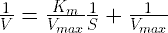
In this way, a straight line is created, providing the user with a much easier way to interpret different quantities and values. Vmax, for instance, is equivalent to the y-intercept of the graph. It is also useful to compare the effect of protein inhibition on Km and Vmax by using the Lineweaver-Burk plot.

Enzyme inhibitors with examples
Almost all life processes are carried out by enzymes. Activation energy is reduced by enzymes when they catalyze a reaction. Enzymes must be tightly controlled, however, to prevent the product from reaching undesired levels. Enzyme inhibition is used to accomplish this.Types
Chemical inhibitors inhibit the activity of enzymes by binding to them. They are reversible and irreversible. They work by immobilizing the enzyme almost permanently. An irreversible inhibitor accomplishes this. There are other chemicals, however, that can temporarily bind to enzymes. Such chemicals are termed reversible. Competitive inhibitors bind to the enzyme's active site (competitive inhibitors), while non-competitive inhibitors bind to other sites on the enzyme.
Competitive inhibitors
The Michaelis-Menten constant (Km) is increased by competitive inhibitors that compete with the substrate at the active site. Despite this, Vmax remains unchanged since the reaction can still be complete with sufficient substrate concentration. When the Km increases, the graph plot of enzyme activity against substrate concentration will shift to the right, whereas the Lineweaver-Burke plot will become steeper when compared to no inhibitor.Non-competitive inhibitors
An inhibitor that binds to a non-competitive site on an enzyme will decrease its VMAX. KM remains the same. Compared with no inhibitor, a graph plotting enzyme activity against substrate concentration demonstrates a lower maximum and a Lineweaver-Burke plot shows a higher y-intercept.
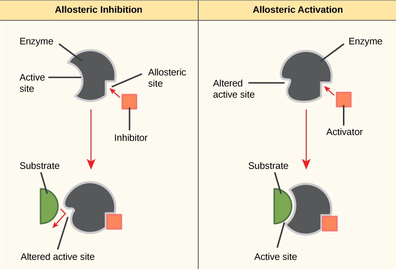
Allosteric inhibitors
An allosteric enzyme displays a sigmoidal curve in contrast to a Michaelis-Menten enzyme, which displays a hyperbolic curve. The reason is that most allosteric enzymes contain multiple subunits that can interact with each other when a substrate is bound to it. K0.5, which is a half-saturation substrate concentration, can be affected by inhibition, or Vmax if it affects both. By reducing Vmax the curve is shifted to the right, and by increasing Vmax the curve is shifted down.The low-affinity state of allosteric enzymes is called the "T" state, while the high-affinity state is called the "R" state. Using inhibitors, an allosteric enzyme maintains its low-affinity state by binding predominantly to its T state. In this way, the amount of enzyme's product can be limited, since it can inhibit the same type of enzyme, preventing excessive amounts of enzyme product from forming. It is referred to as feedback inhibition. ATP allosterically inhibits pyruvate kinase to prevent an increase in pyruvate formation, which results in less ATP being formed. In addition, Kreb's cycle intermediate citrate inhibits phosphofructokinase allosterically. The Kreb's cycle generates a lot of ATP, so glycolysis will be limited when there's a lot of it.
Phosphorylation
An additional mechanism for inhibiting enzymes is phosphorylation. Kinase enzymes primarily play a role in this by either inhibiting or activating enzymes according to the situation. ATP's phosphate group is cleaved off by kinase enzymes, which bind the phosphate group to the enzyme. In situations when this increases enzyme activity, a cascade reaction can be triggered, resulting in a large response from a small stimulus.Zymogens
Zymogens are inactive enzymes secreted in an inactive state. Enzymes are transported safely to a variety of locations by zymogens, but the enzymes do not become active or perform their functions as they travel. Amino acids are added to the proteins so that they remain inactive. To activate a zymogen, this additional amino acid must be removed by another enzyme. Chymotrypsinogen, for example, is synthesized by the pancreas but inactivated and therefore unable to function. In the intestinal tract, an enzyme (trypsin) breaks the additional amino acids into their activated forms (chymotripsin).
Get subject wise printable pdf notesView Here

No comments:
Post a Comment
Please don't spam. Comments having links would not be published.