
Anatomy of urinary tract
Blood is filtered through the urinary system, which creates urine as a waste by-product. The urinary system includes the kidneys, the renal pelvis, the ureters, the bladder, as well as the urethra. As food is digested, nutrients are converted into energy. When the body takes in all the food components it requires, waste products are left in the blood and the bowels. It is the kidneys and the urinary system that assist the body in eliminating liquid wastes such as urea and in maintaining the balance of chemicals such as potassium and sodium. Upon digestion, certain proteins, such as those found in meat, poultry, and certain vegetables, lead to the formation of urea. The urea in the blood is transported to the kidneys, where, together with other wastes, it is removed in the urine.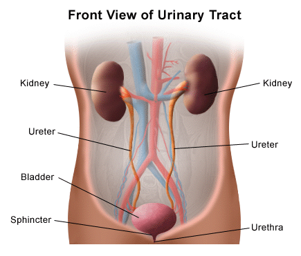
In addition to regulating blood pressure, the kidneys are responsible for producing erythropoietin, which is necessary for red blood cell production in the bone marrow. Other functions of the kidneys include acid-base balance and fluid regulation.
The kidneys and the urinary system: parts and functions
Two kidneys - There are two purplish-brown organs located in the middle of the back, below the ribs. Among their functions are:- Detoxify the body by removing waste products and drugs.
- Ensure that the body's fluids are balanced.
- Regulate blood pressure by releasing hormones.
- Maintain a healthy level of red blood cells.
Two ureters - Both the kidneys and the bladder have these narrow tubes. During excretion, the ureter wall muscles constantly contract and relax, forcing urine downward. Infections in the kidney can occur if urine is allowed to back up or remain stationary. A small amount of urine is excreted from the ureter into the bladder about every 10 to 15 seconds.
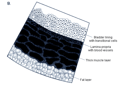
Bladder - The liver is an organ that is in the shape of a triangle located in the lower abdomen. Several ligaments connect the uterus to other organs and pelvic bones. We empty our bladders to accommodate the volume of urine, which relaxes, expands, and flattens the bladder wall. For an extended period of time, an adult bladder can store approximately two cups of urine.

If a bladder abnormality is found during an examination, a specific "landmark" is described. This includes:
- A trigone is a region between the urethra and bladder of the human body
- The lateral wall of the trigone is on either side
- The back wall is the posterior wall
- The dome is the roof of the bladder
Nerves in the bladder - Urination triggers nerves, which alert a person that their bladder needs to be emptied.
Urethra - The tube through which urine leaves the body. Upon receiving the brain's signal, the bladder muscles contract, causing the urine to discharge. A signal is sent from the brain to the sphincter muscles so that urine can be released from the bladder through the urethra. Normally, urination happens when all the signals are present in the correct order.
Anatomy of kidneys
A kidney is one bean-shaped organ that filters minerals from the blood, maintains fluid balance, excretes waste products, and regulates blood volume, among other functions. In the human body, the kidneys play a vital role. The kidneys filter approximately 33% of the blood leaving the heart before pumping it to the body's cells and tissues. In addition to kidney failure, there are often complications involving the kidneys, including fluid retention causing swelling of the extremities, pulmonary Edema (fluid in the lungs), hyperkalaemia (high potassium levels), anaemia, and heart problems.Location
Each kidney is located in the retroperitoneal space on either side of the spine. Due to the liver sitting on the right side of the abdominal cavity, above the right kidney, the left kidney is a little higher than the right one.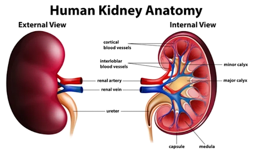
Structure
Bean-shaped organs weigh between 115 grams and 155 grams in females and males. The size of the kidneys is usually 11 to 14 centimeters in length, 6 centimeters wide, and four centimeters in diameter. In the human body, the kidneys are protected by fat, muscles, and ribs. Extrinsic renal fat, also known as the renal fat pad, protects the kidneys from external forces or damage. It plays an important role in providing an entry point and exit point for the vessels, nerves, lymphatics, and ureters associated with the kidney.Vascular supply
An abdominal aorta branch leads to the renal arteries. Specified capillary beds formed by the entry and division of multiple afferent arterioles form the glomeruli. There are several components in a nephron, including the glomerulus. Capillaries merge again after they form afferent arterioles. An outer cortex tubule's efferent arterioles connect to form peritubular networks. As the cortex and medulla approach their inner thirds, the peritubular network is replaced by the vasa recto. Upon leaving the kidneys and entering the heart, filtered blood passes through the left and right renal veins and empties into the inferior vena cava.Parts of kidneys
Anatomy of nephrons
Nephrons in the kidneys produce urine when excess waste and substances are removed from the blood. One million nephrons are found in the human kidney. Among animal embryos, fish, amphibian larva, and early vertebrates often have their earliest nephrons in their kidneys (pronephros). In their early embryonic development, amphibians, fish, and more advanced vertebrates have more advanced nephrons. Vertebrates whose adult kidneys are composed of nephrons are called metanephros, namely reptiles, birds, and mammals. Each nephron of a mammal kidney contains between 30 and 55 mm (1.2 and 2.2 inches) of tubules. An open tube is folded into a double-walled cup at one end, then closed, expanded, and folded again. There are microscopic capillaries called glomeruli in Bowman's capsule, formed from microscopic blood vessels. Renal corpuscles are composed of both a capsule and a glomerulus. A tiny artery called an arteriole travels from and to the glomerulus through the capsule's opening. Filtered fluid from the blood passes through the glomerulus of the kidney and ends up in the nephron tubules through the capsule. Water and other constituents are absorbed into or secreted into filtrates as certain substances are reabsorbed from tubules. Through the collecting tubules, urine is transported to the renal pelvis by the kidneys.Other parts of kidney
The outer layer of the kidney is the cortex, located under the renal capsule. The cortex is lighter in color than the rest of the kidney. There are two groups of convoluted tubules here as well as the renal corpuses. It contains three triangular pieces called renal pyramids, which are located in the medulla, the inner part of the kidney. The collecting ducts are located within the kidney pyramids, in addition to the Loops of Henle. An open apex adorns the kidney pyramid, or renal papilla. In the pyramids, urine is collected by a structure called a minor calyx. When several minor calyces connect, they form a major calyx. Urine passes through the major calices on its way to the renal pelvis. A funnel-shaped structure is formed when the major calices meet at the pelvis. The ureter, which starts at the end of the kidney, receives urine from the kidney at the pelvis-ureteral junction. Gerota's fascia is thin, fibrous, and encloses the adrenal glands above the kidneys. It surrounds the adrenal glands and anchors them to the abdomen. The renal capsule is a tough fibrous layer found within the fat pad of the kidney.
Get subject wise printable pdf notesView Here

No comments:
Post a Comment
Please don't spam. Comments having links would not be published.